Chart Of Nerves In Back
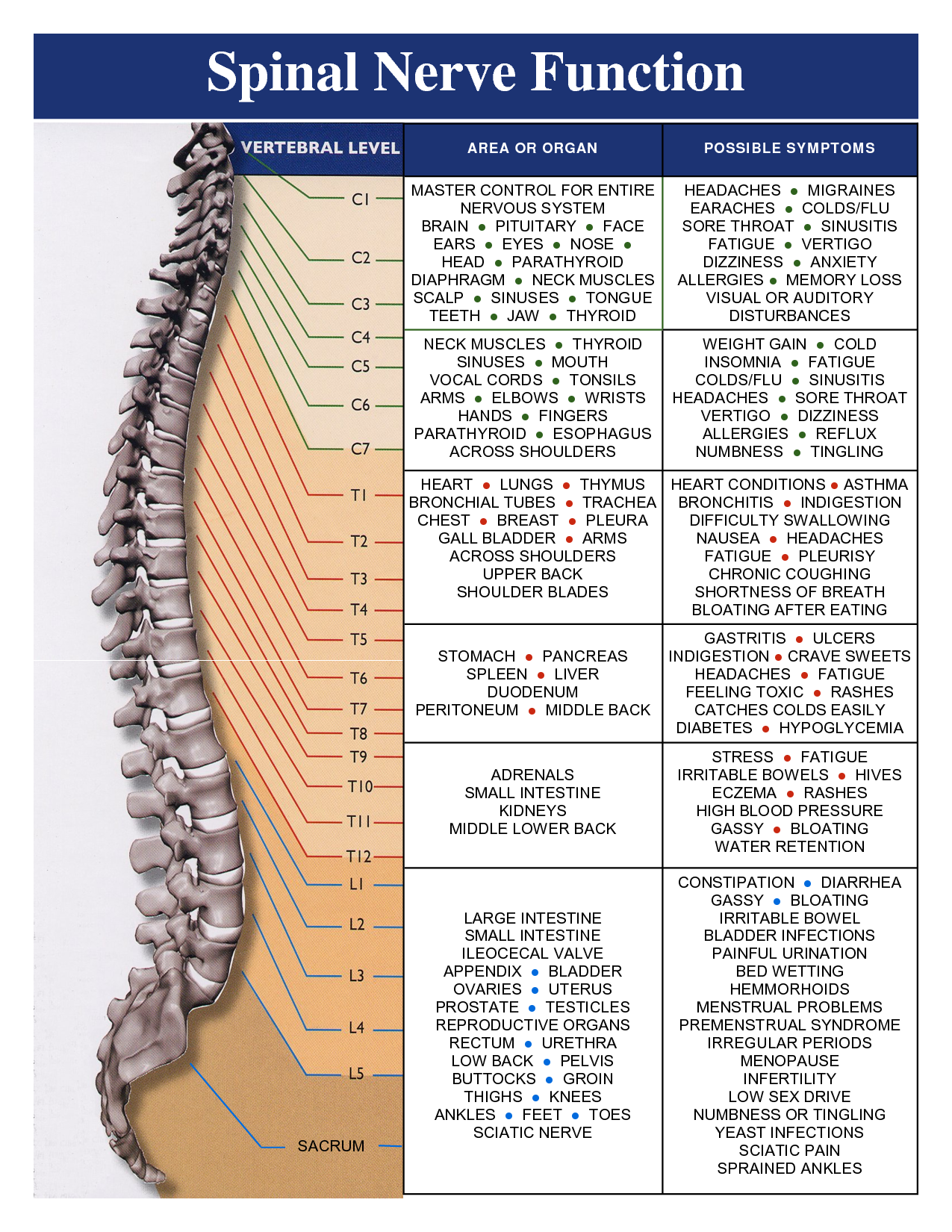
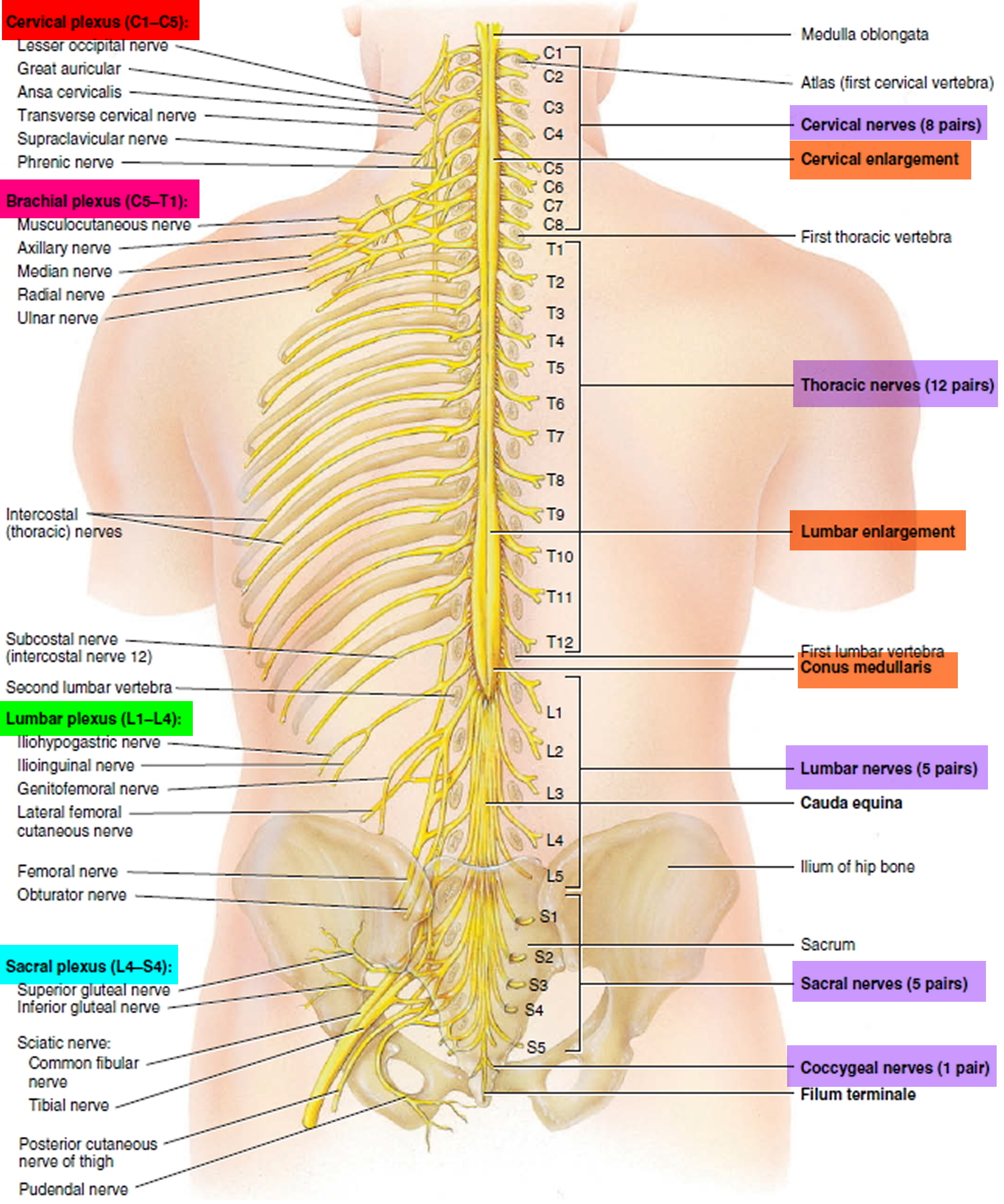
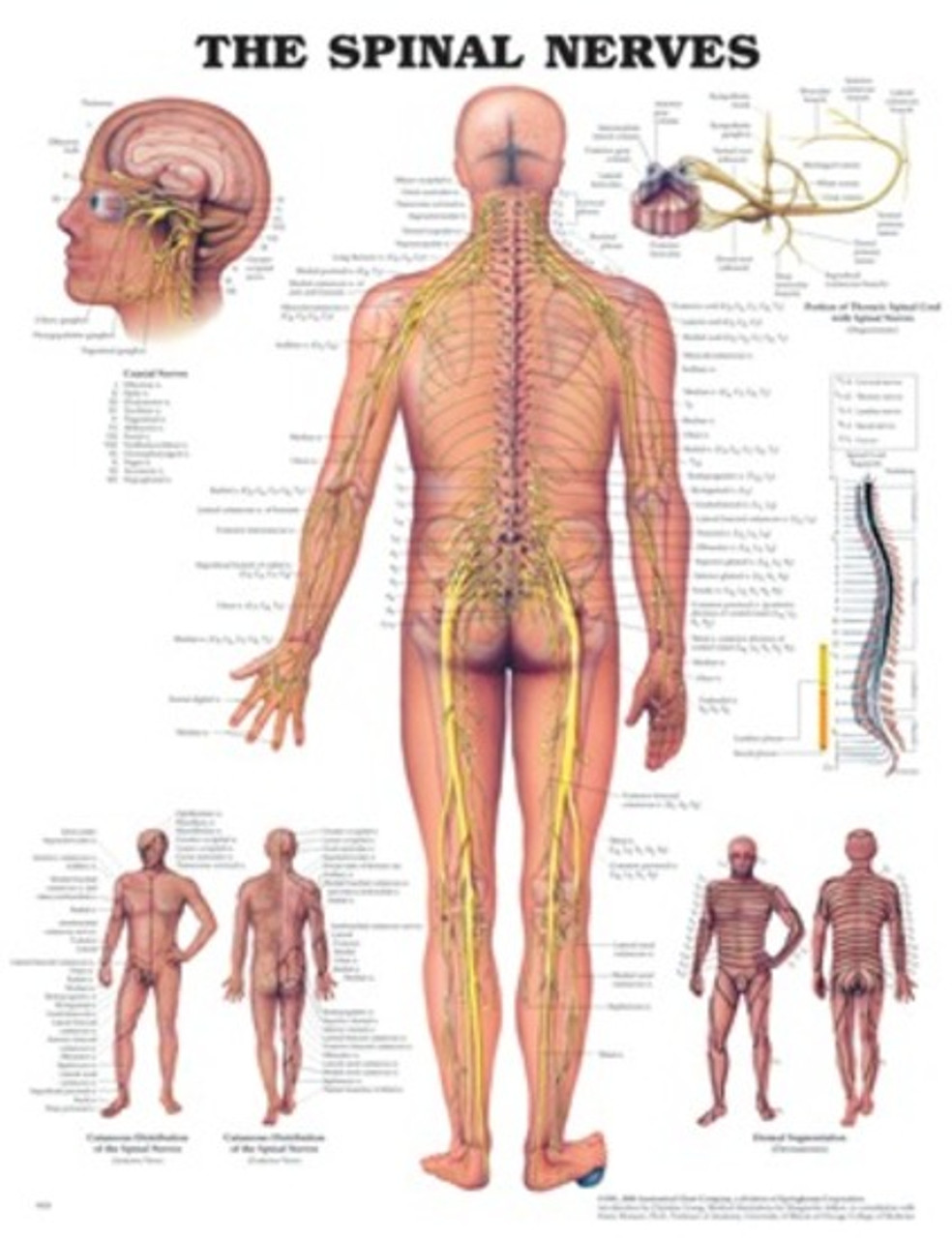
Chart Of Nerves In Back - Seven in your neck (cervical spine ), 12 in your midback (thoracic spine) and 5 in your lower back (lumbar spine). Web is a pinched nerve causing your problem? Sciatica refers to back pain caused by a problem with the sciatic nerve. Web there are seven cervical vertebrae at the top, followed by 11 thoracic vertebrae, five lumbar vertebrae at the lower back, and five fused vertebrae at the bottom to create the sacrum. The lumbar myotomes supply the muscles involved in moving the lower back, hip, knee, foot, and toes. This is a large nerve that runs from the lower back down the back of each leg. Numerous muscles, ligaments and tendons support the spine, providing it with flexibility and a great range of motion. Web how to use the spinal nerve chart: Web spinal nerves are peripheral nerves that emerge from the spinal cord and carry motor, sensory, and autonomic signals between the spinal cord and the rest of the body. Web a dermatome is a distinct area of your skin defined by its connection to one of 30 spinal nerves. The spinal cord begins at the base of the brain and extends into the pelvis. The vertebral column (spine) is the bony core of the back. The peripheral nerves are responsible for sensations and muscle movements. Each pair of spinal nerves are dedicated to certain regions of the body. It is formed by a chain of 33 interconnected vertebrae and their intervening joints. The spinal cord and peripheral nerves. Your spinal cord, made up of billions of nerves, lies inside your spinal column, protected on all sides by bone. When something injures or puts. Web your spinal column or ‘backbone’ is made up of 24 vertebrae: The central system is the primary command center for the body, and is. Web there are 3 basic classes of neurons: Web is a pinched nerve causing your problem? Web in general, the lumbar spinal nerves have dermatomes that receive skin sensations for the parts of the lower back, buttock, thigh, leg, and foot. The lumbar myotomes supply the muscles involved in moving the lower back, hip, knee, foot, and toes. We’ll explore. Web in general, the lumbar spinal nerves have dermatomes that receive skin sensations for the parts of the lower back, buttock, thigh, leg, and foot. Afferent neurons, efferent neurons, and interneurons. Web health library / body systems & organs / spine structure and function. Web these nerves are located at the cervical (neck), thoracic (upper back), lumbar (lower back), sacral. Web how to use the spinal nerve chart: The peripheral nerves are responsible for sensations and muscle movements. Afferent neurons, efferent neurons, and interneurons. Spinal nerves are an integral part of the peripheral nervous system (pns). They are the structures through which the central nervous system (cns) receives sensory information from the periphery, and through which the activity of the. Many of the nerves of the peripheral nervous system, or pns, branch out from the spinal cord and travel to. Web this is a dynamic list and may never be able to satisfy particular standards for completeness. The spinal cord and peripheral nerves. Each pair of spinal nerves are dedicated to certain regions of the body. The vertebral column (spine). The spinal cord and peripheral nerves. Web how to use the spinal nerve chart: Also known as sensory neurons, afferent neurons transmit sensory signals to the central nervous system from receptors in the body. The spinal cord begins at the base of the brain and extends into the pelvis. This is a large nerve that runs from the lower back. Lists of human anatomical features. The lumbar myotomes supply the muscles involved in moving the lower back, hip, knee, foot, and toes. Your spine is made up of vertebrae (bones), disks, joints, soft tissues, nerves and your spinal cord. 8 cervical, 12 thoracic, 5 lumbar, 5 sacral, and 1 coccygeal, named according to their corresponding vertebral levels. Also known as. 8 cervical, 12 thoracic, 5 lumbar, 5 sacral, and 1 coccygeal, named according to their corresponding vertebral levels. Web the nervous system has two major parts: Web a dermatome is a distinct area of your skin defined by its connection to one of 30 spinal nerves. Numerous muscles, ligaments and tendons support the spine, providing it with flexibility and a. The lumbar myotomes supply the muscles involved in moving the lower back, hip, knee, foot, and toes. Web learn how spinal nerve roots function, and the potential symptoms of spinal nerve compression and pain in the neck and lower back. Web these nerves are located at the cervical (neck), thoracic (upper back), lumbar (lower back), sacral (sacrum, which forms part. Your spine is made up of vertebrae (bones), disks, joints, soft tissues, nerves and your spinal cord. The lumbar myotomes supply the muscles involved in moving the lower back, hip, knee, foot, and toes. Many of the nerves of the peripheral nervous system, or pns, branch out from the spinal cord and travel to. Afferent neurons, efferent neurons, and interneurons.. The spinal cord begins at the base of the brain and extends into the pelvis. Web these nerves are located at the cervical (neck), thoracic (upper back), lumbar (lower back), sacral (sacrum, which forms part of the pelvis), and coccygeal (tailbone) levels. Web how to use the spinal nerve chart: The lumbar myotomes supply the muscles involved in moving the. The spine is composed of. Web is a pinched nerve causing your problem? The back comprises the spine and spinal nerves, as well as several different muscle groups. Also known as sensory neurons, afferent neurons transmit sensory signals to the central nervous system from receptors in the body. Your spine is an important bone structure that supports your body and helps you walk, twist and move. Web this spinal nerve pain chart provides a pictorial representation of three types of nerves: Web these nerves are located at the cervical (neck), thoracic (upper back), lumbar (lower back), sacral (sacrum, which forms part of the pelvis), and coccygeal (tailbone) levels. We’ll explore more about both your spinal nerves and dermatomes, including a chart showing. This is a large nerve that runs from the lower back down the back of each leg. The vertebral column (spine) is the bony core of the back. Web there are 3 basic classes of neurons: The central system is the primary command center for the body, and is. You can help by adding missing items with reliable sources. It is formed by a chain of 33 interconnected vertebrae and their intervening joints. 8 cervical, 12 thoracic, 5 lumbar, 5 sacral, and 1 coccygeal, named according to their corresponding vertebral levels. When something injures or puts.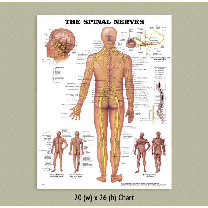
Back Talk Systems, Colorado »Spinal Nerves Anatomical Chart
Vital Connections Briggs Chiropractic Clinic
Spinal Cord Anatomy Parts and Spinal Cord Functions
spinalnervechart Schertz Chiropractic

Nerve chart Subluxation, Chiropractic care, Spine health

Spinal Nerve Chart

Spinal Nerve Chart Print 5x7 Etsy Nerve anatomy, Spinal nerves

Diagram Of Nerves In Lower Back

All About The Spinal Cord Spinal Nerve Distribution C vrogue.co
Spinal Nerves Anatomical Chart Southern Biological
Your Spinal Cord, Made Up Of Billions Of Nerves, Lies Inside Your Spinal Column, Protected On All Sides By Bone.
The Peripheral Nerves Are Responsible For Sensations And Muscle Movements.
The Central Nervous System (Cns) And The Peripheral Nervous System (Pns).
It Forms The Axial Skeleton Together With The Skull And Rib Cage.
Related Post: